 |
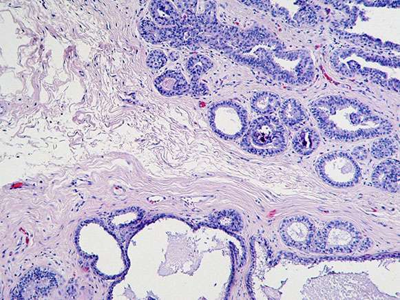 |
| Figure 1. H&E, 10x. | Figure 2. H&E, 10x. |
 |
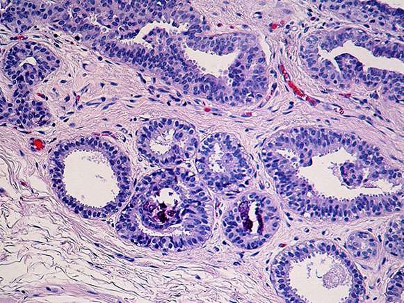 |
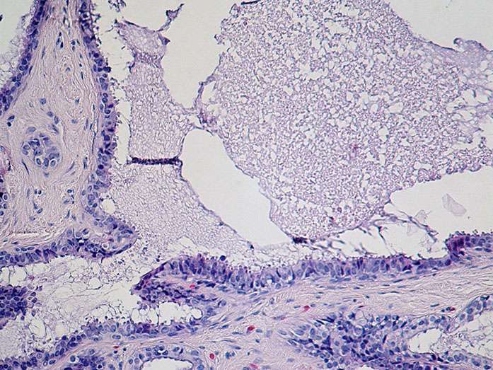
| Figure 3. H&E, 20x. (columnar cell hypeplasia with focal atypia/FEA) | Figure 4. H&E, 20x. (columnar cell lesion with calcifications) |
 |
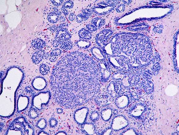 |
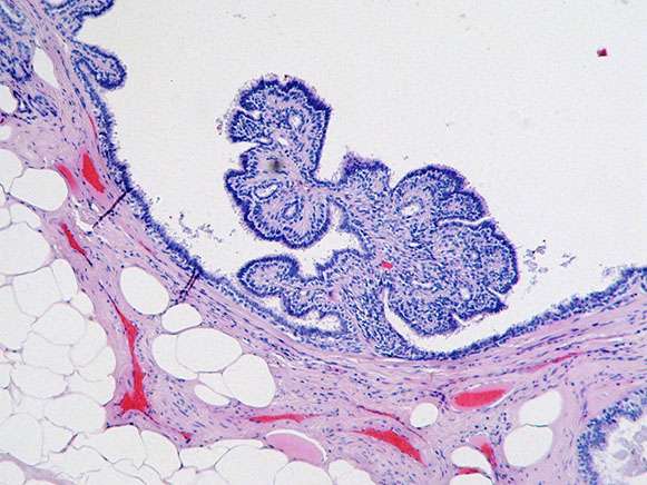
| Figure 5. H&E, 10x. (intraductal papilloma) | Figure 6. Fibrocystic changes with florid ductal hyperplasia |
