 |
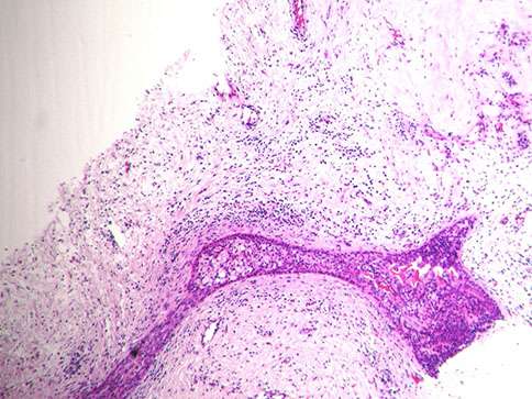 |
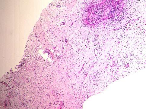
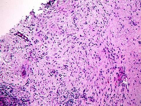
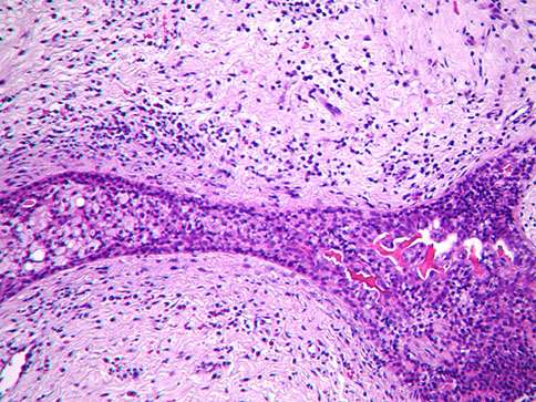
| Figure 1. H&E, 5x. | Figure 2. H&E, 5x. (note the inflammatory response within the myxoid stroma and residual DCIS) |
 |
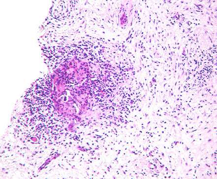 |
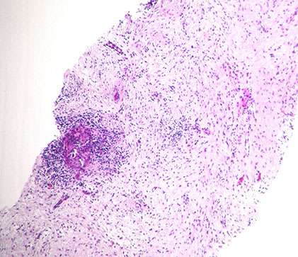
| Figure 3. H&E, 5x. | Figure 4. H&E, 10x. |
 |
 |
| Figure 5. H&E, 10x. | Figure 6. H&E, 20x. |
 |
|
| Figure . H&E, 20x. |

Copyright 2012 – 2024 | BosnianPathology.org | Semir Vranic MD, PhD | Faruk Skenderi MD, PhD
next generation pathology